
Code for imaging. AI models that keep working in the clinic.
What you see here is a CT scan of a patient with a suspected stroke. We build the AI model that analyzes a scan like this. And not only CT: also MRI, ultrasound and pathology slides.
A model learns from examples: images that an expert has annotated by hand. From collecting those images to running the model in the clinic, it is all software. So a model is only as good as the engineering behind it. That is what we do.
For you, that means a product that works. A model that is right 95% of the time leaves one case in twenty for someone to correct. At 99.9%, it is one in a thousand. Your customers work faster and trust the result, and a product they trust is a product that sells.
An AI model is never finished. When the scanners, the clinical insights or the patient populations change, the data drifts away from what the model has learned, and its performance degrades. So a model has to be monitored and updated, and sometimes rebuilt. AI development in this domain never stops.
Trusted by
Xyall
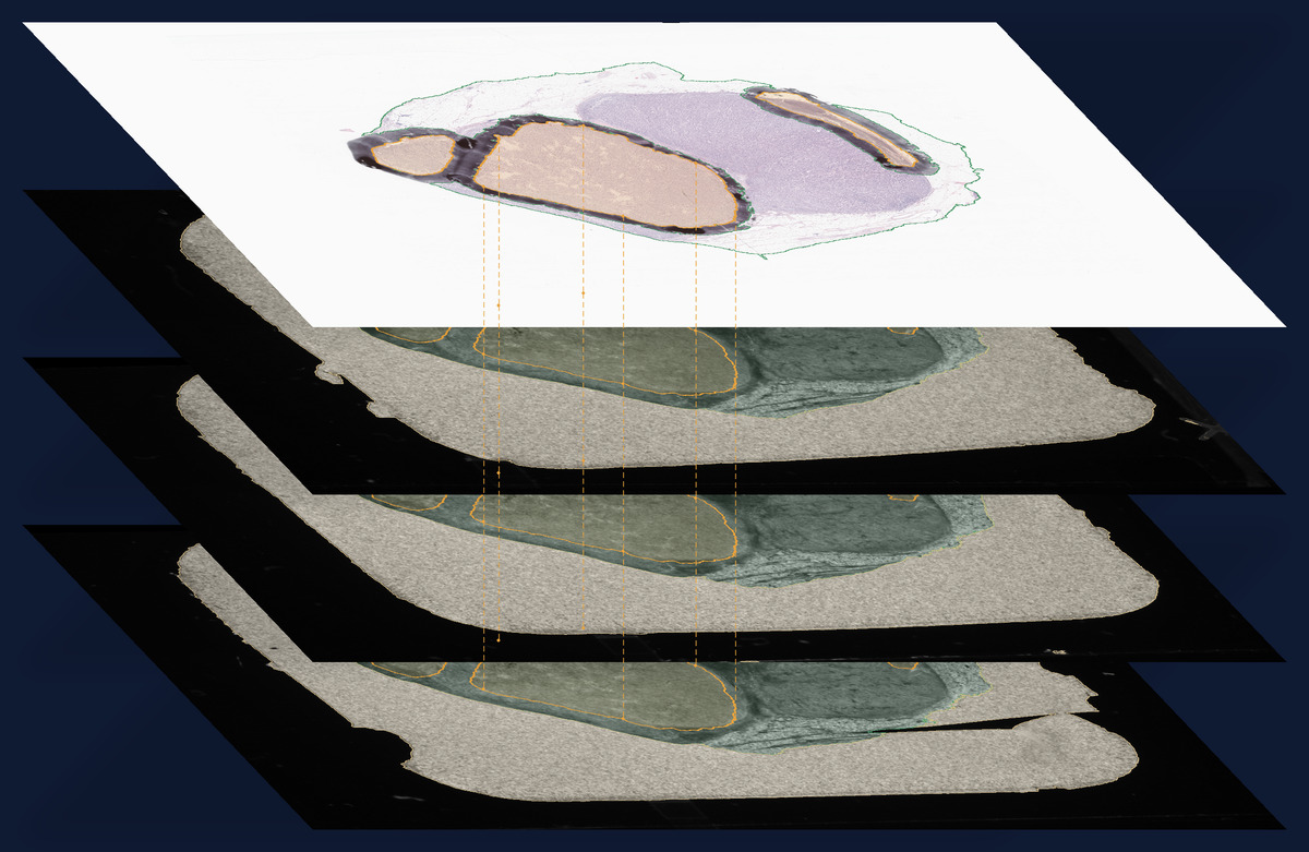
Slide registration for a tissue-collecting robot
Canon
Vessel segmentation and collateral scoring in CTA
Radboudumc
The first brain tissue segmentation in 4D CTA
How we build
Superstack
Our AI development environment
DaVinci
Our cloud-based annotation platform
In production
Rashindra’s sharp mind makes working with him a delight. Rather than relying on off-the-shelf tools, he challenged us to truly understand our data. XDMD’s approach led to a custom solution that significantly improved our image registration process.
Senior Software Architect at Xyall, The Netherlands
How we work
Understand the problem first
We find out what the problem really is, and why things are as they are. Then we zoom out and ask whether it is the right problem to solve, before we write a single line of code.
The best code is no code
What we do not build cannot fail and needs no maintenance. What we build is only what the job needs.
Never fail silently
When a case is outside what our software can handle, it says so and stops, instead of quietly giving a wrong result.
Watch every model in use
Scanners, protocols and patients change. We check each new case against what the model learned from, and update the model before its results degrade.
Who we are
Our team and advisors have spent decades in academic hospitals and in industry, and have written more than 1000 peer-reviewed publications. We built this expertise before and during the rise of AI, and today we use AI to build AI.
Rashindra is one of the most knowledgeable engineers in medical imaging and machine learning. If you look for solid solutions for your image analysis problems, look no further.
Professor of Medical Image Analysis at Radboudumc, Founder at Plain Medical, Founder at Thirona, The Netherlands
Work with us
We work with companies that build products with medical images, and with hospitals and research groups. Tell us in a few lines what the problem is and what data you have. Within one week we tell you how we would approach it. If we go ahead: a pilot on your data first, then the model in production, and we keep it working.